Work
Professional projects across drug discovery, medical imaging, and infrastructure.
§ AstraZeneca · 2019 – present
Three threads: drug discovery ML, medical imaging and segmentation, and the tooling and infrastructure that supports both. Grouped by thread, most recent work first inside each.
Drug Discovery ML
Multimodal IC50 prediction
PyTorch models fusing cell painting microscopy with SMILES chemical representations for compound activity prediction. Deployed across compound libraries totaling 39K compounds; jointly learned image-and-structure embeddings inform downstream phenotype-to-target reasoning.Graph-based analysis of multiplex immunofluorescence
Graph Neural Networks over multi-channel images, capturing spatial cell-cell relationships in multiplex IF for downstream phenotype classification. Preprint on bioRxiv; abstracts at SITC and AACR.Medical Imaging & Segmentation
Automatic contrast phase classification of polyphasic CT scans
Deep learning classifier inferring contrast phase from CT volumes — a prerequisite for downstream tumor segmentation pipelines that depend on consistent imaging protocol. Co-authored work, poster at AACR 2026.Interactive 3D segmentation toolset
Transformer-backed segmentation tool for 3D volumetric data with text-guided prompts. Halved annotation time and deployed to twelve internal users across R&D. Companion paper as poster at AACR 2024.Tooling & Infrastructure
Biomedical imaging data platform
Unified ingestion and preprocessing platform for biomedical imaging at scale — now the team’s standard data layer. Sixteen multi-center datasets, 150K CT volumes, automated mapping of thousands of annotation masks across DICOM and NIfTI standards.Embedding visualization toolkit
Web-based visualization suite for high-dimensional embedding interpretation — clustering (HDBSCAN over UMAP), heatmaps, histograms, archetype analysis. Used by R&D labs across several modeling projects.§ Ann Arbor Algorithms · 2018 – 2019
Software Engineer building containerized end-to-end deep learning pipelines for medical imaging — classification, 3D bounding-box detection, and anomaly identification across multimodal datasets at scale.
Microcalcification detection in mammography
End-to-end deep learning detection of microcalcifications using a U-Net architecture, with downstream localization of asymmetric patterns. The work was peer-reviewed and published in Patterns — Guan, Wang, Li, Zhang, Chen, Siddiqui, Nehring, Huang. Detecting asymmetric patterns and localizing cancers on mammograms. Patterns 1, no. 7 (2020).Other work at AAA:
- Chest vessel segmentation — 3D deep learning for atherosclerosis identification on chest MRI.
- Integrated tumor segmentation — Dockerized lung-cancer segmentation with 3D visualization.
- Lung cancer prediction — XGBoost over patient metadata (~1.6K patients).
- ECG abnormality identification — ResNet variant for 12-lead ECG analysis (~7K samples).
- Colorectal surgical phase detection — video and sensor-based phase prediction.
Demos
AZ work is largely confidential. These demos from earlier work at Ann Arbor Algorithms remain publicly viewable.
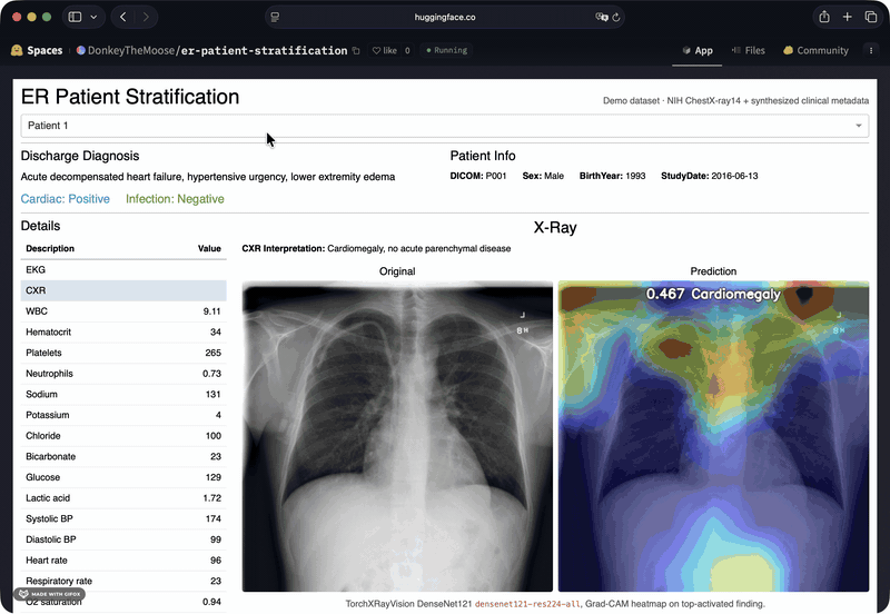
Patient Info — disease prediction dashboard
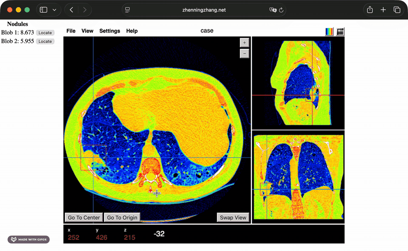
Papaya — lung-nodule DICOM viewer
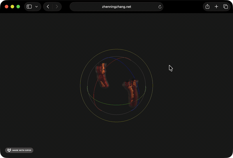
Plaque — chest-vessel atherosclerosis 3D viewer
For personal projects and side builds, see Tinkering.